
Fil:Fundus of patient with retinitis pigmentosa, mid stage.jpg
Størrelse af denne forhåndsvisning: 699 × 599 pixels. Andre opløsninger: 280 × 240 pixels | 560 × 480 pixels | 871 × 747 pixels.
Fuld opløsning (871 × 747 billedpunkter, filstørrelse: 107 KB, MIME-type: image/jpeg)
|
|
Denne fil er fra Wikimedia Commons. Beskrivelsen af filen fra Commons er gengivet nedenfor. |
| Beskrivelse |
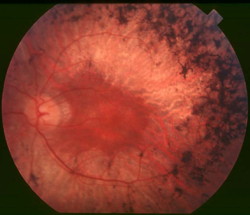
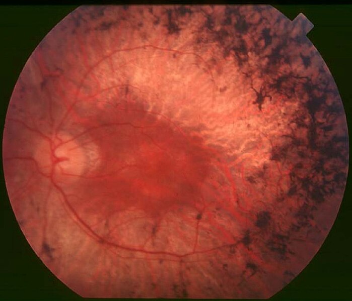
English: Figure 2. Fundus of patient with retinitis pigmentosa, mid stage (Bone spicule-shaped pigment deposits are present in the mid periphery along with retinal atrophy, while the macula is preserved although with a peripheral ring of depigmentation. Retinal vessels are attenuated.) Hamel Orphanet Journal of Rare Diseases 2006 1:40 doi:10.1186/1750-1172-1-40 |
| Dato | |
| Kilde | Retinitis pigmentosa by Christian Hamel |
| Forfatter | Christian Hamel |
| Tilladelse (Genbrug af denne fil) |
© 2006 Hamel; licensee BioMed Central Ltd. This is an Open Access article distributed under the terms of the Creative Commons Attribution License (https://creativecommons.org/licenses/by/2.0), which permits unrestricted use, distribution, and reproduction in any medium, provided the original work is properly cited. |
Denne fil er udgivet under Creative Commons Navngivelse 2.0 Generisk-licensen
- Du må frit:
- at dele – at kopiere, distribuere og overføre værket
- at remixe – at tilpasse værket
- Under følgende vilkår:
- kreditering – Du skal give passende kreditering, angive et link til licensen, og oplyse om der er foretaget ændringer. Du må gøre det på enhver fornuftig måde, men ikke på en måde der antyder at licensgiveren godkender dig eller din anvendelse.
Filhistorik
Klik på en dato/tid for at se filen som den så ud på det tidspunkt.
| Dato/tid | Miniaturebillede | Dimensioner | Bruger | Kommentar | |
|---|---|---|---|---|---|
| nuværende | 2. dec. 2017, 12:17 | | 871 × 747 (107 KB) | Doc James | Cropped 27 % horizontally and 7 % vertically using CropTool with precise mode. |
| 22. sep. 2009, 15:52 |  | 1.200 × 799 (126 KB) | CopperKettle | {{Information |Description={{en|1=Figure 2. Fundus of patient with retinitis pigmentosa, mid stage (Bone spicule-shaped pigment deposits are present in the mid periphery along with retinal atrophy, while the macula is preserved although with a peripheral |
Filanvendelse
Den følgende side bruger denne fil:
Global filanvendelse
Følgende andre wikier anvender denne fil:
- Anvendelser på ar.wikipedia.org
- Anvendelser på bs.wikipedia.org
- Anvendelser på ca.wikipedia.org
- Anvendelser på en.wikipedia.org
- Anvendelser på en.wikiversity.org
- Anvendelser på es.wikipedia.org
- Anvendelser på eu.wikipedia.org
- Anvendelser på fa.wikipedia.org
- Anvendelser på fi.wikipedia.org
- Anvendelser på fr.wikipedia.org
- Anvendelser på he.wikipedia.org
- Anvendelser på hy.wikipedia.org
- Anvendelser på it.wikipedia.org
- Anvendelser på ko.wikipedia.org
- Anvendelser på la.wikipedia.org
- Anvendelser på or.wikipedia.org
- Anvendelser på outreach.wikimedia.org
- Anvendelser på pl.wikipedia.org
- Anvendelser på pt.wikipedia.org
- Anvendelser på ru.wikipedia.org
- Anvendelser på sl.wikipedia.org
- Anvendelser på sr.wikipedia.org
- Anvendelser på sv.wikipedia.org
- Anvendelser på th.wikipedia.org
- Anvendelser på tr.wikipedia.org
- Anvendelser på tt.wikipedia.org
- Anvendelser på uk.wikipedia.org
- Anvendelser på vi.wikipedia.org
Vis flere globale anvendelser af denne fil.